Universal UltraSound Image Challenge: Multi-Organ Classification and Segmentation

A challenge accepted by MICCAI 2025, 23-27 September, Daejeon, Republic of Korea.

Participate Learn more
Ultrasound Image Analysis
Ultrasound imaging is a vital tool in biomedical diagnostics, widely used for organ examination and tumor detection.
Generalized Learning
Universal models require advanced learning techniques to handle diverse organs and pathologies.
Multi-task Learning
Multi-task learning can aid in precise diagnoses and improve clinical outcomes.

Aim of the challenge

Ultrasound imaging is a vital diagnostic tool, but accurately analyzing images across diverse organs and pathologies remains challenging. Existing methods often lack generalization and are limited to specific tasks.
  • Developing a Universal Model
    To design a generalized model capable of handling multiple tasks in ultrasound image analysis, including classification and segmentation across diverse organs and pathologies.
  • Advancing Machine Learning Techniques
    To explore and implement novel machine learning and image processing methods for extracting meaningful features and achieving accurate predictions.
  • Improving Clinical Outcomes
    To enhance diagnostic accuracy and efficiency, potentially reducing invasive procedures and improving patient outcomes through better ultrasound image analysis.
responsive devices

Task

Participants are required to submit a Docker image containing a single model capable of performing multi-task processing across multiple organs and diseases. The target organs include the breast, thyroid, liver, kidney, fetal head, heart, and appendix, which correspond to publicly available datasets we collected for redistribution.

Specifically:
  • Breast: BUSI, BUSIS, BUS-BRA datasets (tasks: nodule malignancy classification + nodule segmentation)
  • Thyroid: DDTI dataset (task: nodule segmentation)
  • Liver: Fatty-Liver dataset (task: fatty liver classification)
  • Kidney: KidneyUS dataset (task: kidney contour delineation)
  • Fetal Head: Fetal HC dataset (task: fetal head contour segmentation)
  • Cardiac: CAMUS dataset (task: cardiac contour segmentation)
  • Appendix: Appendix dataset (task: appendicitis classification)
  • Each organ is associated with a distinct clinical task, exemplifying the multi-disease and multi-task nature of the challenge. In addition to the public datasets, we will provide a small portion of our private datasets for training to mitigate domain gap difficulties. The validation and test sets will consist primarily of private data, which will not be publicly disclosed.

    Data

    Our dataset is combined from multiple public datasets and several private datasets for ultrasound imaging. For the public datasets, detailed information about the data-acquiring centers or institutes can be found in the corresponding articles we have listed. Regarding the private data sources, they are from three hospitals: Zhejiang Cancer Hospital, Hangzhou First People's Hospital, and the Netherlands Cancer Institute.

    Data annotation is overseen by three experts in the field of ultrasound: Dr. Lingyun Bao, Dr. Dong Xu, and Dr. Ritse Mann. They have extensive knowledge and hands-on experience in ultrasound imaging and diagnosis.

    References
    [1] Zehui Lin, Zhuoneng Zhang, Xindi Hu, Zhifan Gao, Xin Yang, Yue Sun, Dong Ni and Tao Tan. "UniUSNet: A Promptable Framework for Universal Ultrasound Disease Prediction and Tissue Segmentation" In 2024 IEEE International Conference on Bioinformatics and Biomedicine (BIBM), IEEE, 2024.
    [2] Tianyu Zhang, Tao Tan, Luyi Han, Linda Appelman, Jeroen Veltman, Ronni Wessels, Katya Duvivier, Claudette Loo, Yuan Gao, Xin Wang, Hugo Horlings, Regina Beets-Tan, Ritse Mann. "Predicting breast cancer types on and beyond molecular level in a multi-modal fashion." NPJ Breast Cancer 9, no. 1 (2023): 16.
    [3] Al-Dhabyani, Walid, Mohammed Gomaa, Hussien Khaled, and Aly Fahmy. "Dataset of breast ultrasound images." Data in brief 28 (2020): 104863.
    [4] Zhang, Yingtao, Min Xian, Heng-Da Cheng, Bryar Shareef, Jianrui Ding, Fei Xu, Kuan Huang, Boyu Zhang, Chunping Ning, and Ying Wang. "BUSIS: a benchmark for breast ultrasound image segmentation." In Healthcare, vol. 10, no. 4, p. 729. MDPI, 2022.
    [5] Gómez-Flores, Wilfrido, Maria Julia Gregorio-Calas, and Wagner Coelho de Albuquerque Pereira. "BUS-BRA: A breast ultrasound dataset for assessing computer-aided diagnosis systems." Medical Physics 51, no. 4 (2024): 3110-3123.
    [6] Byra, Michał, Grzegorz Styczynski, Cezary Szmigielski, Piotr Kalinowski, Łukasz Michałowski, Rafał Paluszkiewicz, Bogna Ziarkiewicz-Wróblewska, Krzysztof Zieniewicz, Piotr Sobieraj, and Andrzej Nowicki. "Transfer learning with deep convolutional neural network for liver steatosis assessment in ultrasound images." International journal of computer assisted radiology and surgery 13 (2018): 1895-1903.
    [7] Singla, Rohit, Cailin Ringstrom, Grace Hu, Victoria Lessoway, Janice Reid, Christopher Nguan, and Robert Rohling. "The open kidney ultrasound data set." In International Workshop on Advances in Simplifying Medical Ultrasound, pp. 155-164. Cham: Springer Nature Switzerland, 2023.
    [8] Pedraza, Lina, Carlos Vargas, Fabián Narváez, Oscar Durán, Emma Muñoz, and Eduardo Romero. "An open access thyroid ultrasound image database." In 10th International symposium on medical information processing and analysis, vol. 9287, pp. 188-193. SPIE, 2015.
    [9] van den Heuvel, Thomas LA, Dagmar de Bruijn, Chris L. de Korte, and Bram van Ginneken. "Automated measurement of fetal head circumference using 2D ultrasound images." PloS one 13, no. 8 (2018): e0200412.
    [10] Leclerc, Sarah, Erik Smistad, Joao Pedrosa, Andreas Østvik, Frederic Cervenansky, Florian Espinosa, Torvald Espeland et al. "Deep learning for segmentation using an open large-scale dataset in 2D echocardiography." IEEE transactions on medical imaging 38, no. 9 (2019): 2198-2210.
    [11] Marcinkevičs, Ričards, Patricia Reis Wolfertstetter, Ugne Klimiene, Ece Özkan Elsen, Kieran Chin-Cheong, Alyssia Paschke, Julia Zerres et al. "Regensburg pediatric appendicitis dataset."

    Rules

    Methods
  • Participants are allowed to base their methods on existing open-source general models. And they are invited to submit a short paper to the Deep-Breath workshop to introduce the details of their methods and declare innovations in areas such as preprocessing, model architecture, or post-processing.
  • Use of other training data/pre-trained models
  • No additional data allowed;
  • The publicly available pre-trained model, such as ResNet50 trained on ImageNet, can be used. To ensure fairness and reproducibility of results, using unpublished pre-trained models is prohibited.
  • Award Eligibility

    As a condition for being ranked and considered as the challenge winner or eligible for any prize, the teams/participants must fulfil the following obligations:

  • Present their method at the final event of the challenge at MICCAI 2025;
  • Submit a paper to Deep-Breath workshop reporting the details of the methods in a short or long (up to the teams) LNCS format;
  • Sign and return all prize acceptance documents as may be required by Competition Sponsor/Organizers;
  • Commit to citing the data challenge paper and the data overview paper whenever submitting the developed method for scientific and non-scientific publications.
  • Evaluation

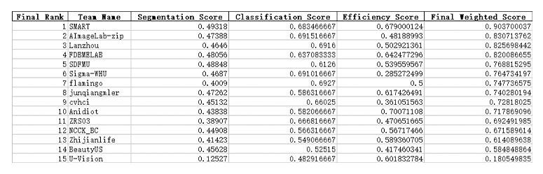
    The algorithm will be assessed using the following metrics, and all metrics will be used to compute the ranking.

    Segmentation Metrics
  • Dice Similarity Coefficient (DSC): Measures overlap between predicted and ground truth segmentations.
  • Normalized Surface Dice (NSD): Evaluates boundary accuracy within a tolerance distance.
  • Classification Metrics
  • Area Under Curve (AUC): Assesses classification performance across thresholds.
  • Accuracy: Proportion of correctly classified instances.
  • Efficiency Metrics
  • Running Time: Inference time for computational efficiency.
  • Maximum GPU Memory: Peak memory usage during inference.
  • Timeline

    • July 20, 2025 Website opens for registration, release training and validation images.
    • Aug. 20, 2025 Submission system opens for validation.
    • Sept. 1, 2025 Submission system opens for testing.
    • Sept. 15, 2025 Registration and docker submission deadline.
    • Sept. 23-27, 2025 Release final results during the MICCAI annual meeting.

    Awards

    • Top-10 teams will be invited to present talks and winning certificates will be provided.
    • We will provide a prize for the Top 3 teams. The exact money of cash prize is not known yet.
    • Sponsors for the awards are currently being investigated.
    • The challenge organizers aim to publish a summary paper in a relevant journal. The top ten performing teams will be invited to as co-authors. Participants can submit their own paper after submitting the summary paper or once the challenge paper is published on Arxiv.

    UUSIC Committee

    Organizing Committee

    Zehui Lin

    Faculty of Applied Sciences, Macao Polytechnic University, Macao, China

    Luyi Han

    Radiology Department/BIG, NKI/RadboudUMC, Amsterdam/Nijmegen, the Netherlands

    Tianyu Zhang

    Radiology Department/BIG, NKI/RadboudUMC, Amsterdam/Nijmegen, the Netherlands

    Xing Wang

    Radiology Department/BIG, NKI/RadboudUMC, Amsterdam/Nijmegen, the Netherlands

    Shuo Li

    School of Biomedical Engineering, Case Western Reserve University, Cleveland, USA

    Shandong Wu

    Department of Radiology, University of Pittsburgh, Pittsburgh, USA

    Dong Ni

    School of Biomedical Engineering, Shenzhen University, China

    Dong Xu (Co-chair)

    Department of Diagnostic Ultrasound Imaging & Interventional Therapy, Zhejiang Cancer Hospital, Hangzhou Institute of Medicine (HIM), Chinese Academy of Sciences, Hangzhou, China

    Tao Tan (Chair)

    Faculty of Applied Sciences, Macao Polytechnic University, Macao, China

    Technical Committee

    Oliver Lester Saldanha

    NCT in Uniklinik Heidelberg and EKFZ in TU Dresden, Dresden, Saxony, Germany

    Stefano Trebeschi

    Radiology Department, Netherlands Cancer Institute
    GROW Graduate School, Maastricht University, Netherlands

    Ehsan Kozegar

    Faculty of Technology and Engineering-East of Guilan, University of Guilan, Rudsar, Guilan, Iran

    Dengqiang Jia

    City University of Hong Kong, Hong Kong, China

    Yapeng Wang

    Faculty of Applied Sciences, Macao Polytechnic University, Macao, China

    Gustav Müller-Franzes

    Department of Diagnostic and Interventional Radiology, University Hospital RWTH Aachen, Aachen, Germany

    Behdad Dasht Bozorg

    Image-Guided Surgery, Department of Surgery, NKI, Amsterdam, the Netherlands

    Clinical Committee

    Lingyun Bao

    Affiliated Hangzhou First People's Hospital, Zhejiang University School of Medicine, Hangzhou, China

    Jieyun Bai

    Jinan University, China

    Ying Zhou

    Faculty of Applied Sciences, Macao Polytechnic University, Macao, China
    Department of Surgery, Hebei Provincial Hospital of Traditional Chinese Medicine, Shijiazhuang, China

    Yanming Zhang

    Faculty of Applied Sciences, Macao Polytechnic University, Macao, China

    Ritse Mann

    Radiology Department/BIG, NKI/RadboudUMC, Amsterdam/Nijmegen, the Netherlands

    Contact

    Please contact us for further questions and comments via email at uusic2025@gmail.com

    Sponsors